Science Summary For Class 9 Tissues

Science Summary For Class 9 Tissues

Tissue:- A Term coined by Bichat (1771-1802). Bichat distinguished 21 elementary tissues from which the organs of the Human body are composed. 

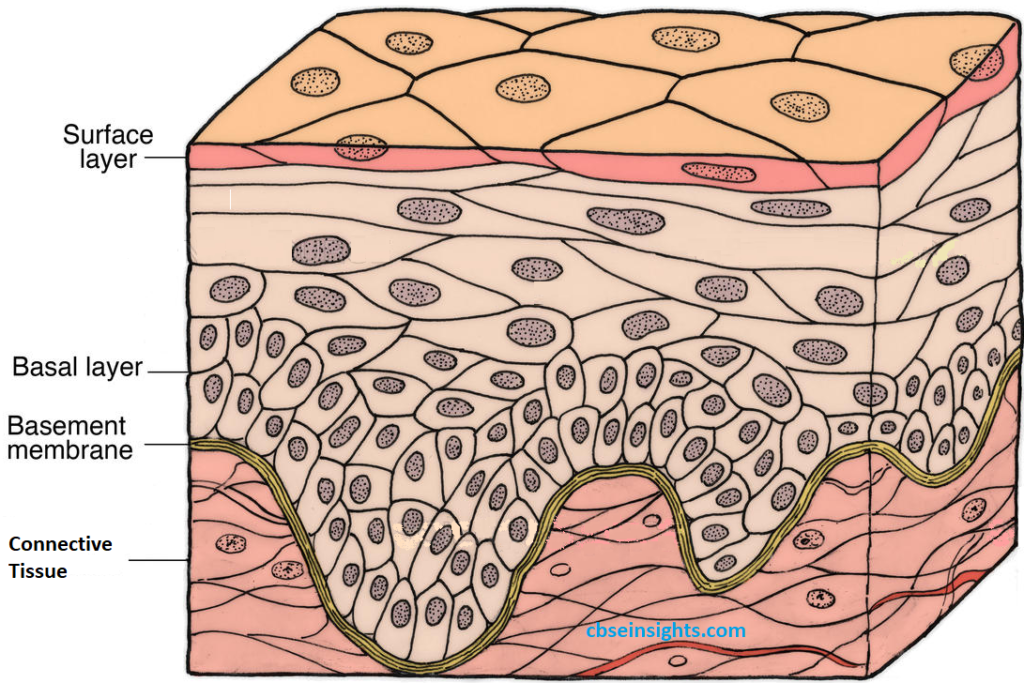
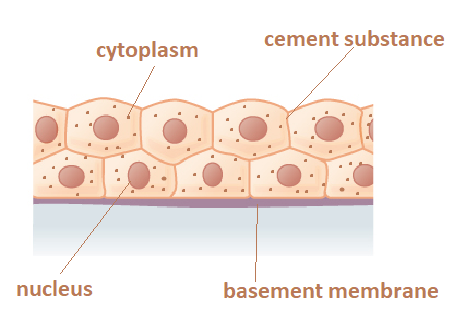
A group of cells that are specialized to perform a particular function forms a tissue.

Importance of Tissues :- 

  1.  Due to the formation of tissues , Division of Labour is possible in the Multicellular Organisms. 
  2. Tissues form Organs , Organs form Organ Systems.
  3. Individual cell is now less burdened due to Division of Labour. 
  4. The efficiency and the chances of Survival increases in the Multicellular Organisms. 

Tissues are mainly classified into two types:

1. Plant Tissues

2. Animal Tissues

1.Plant tissues

  1.  Plants do not move, i.e., they are stationary.
  2. Most of the tissues they have are supportive, which provides them with structural strength.
  3.  Most of these tissues are dead, as they can provide better mechanical strength than the live ones, and need less maintenance.
  4. Some of the plant tissues keep on dividing throughout the plant life. These tissues are localised in certain regions.

Types of Plant Tissues:

Based on the dividing capacity of the tissues, various plant tissues can be classified as (growing or meristematic tissue) and permanent tissue which have further sub-divisions as explained below:

Tissues

Group of cells having a common origin and similar function are termed as tissues.


A. Plant tissues: On the basis of the dividing capacity, plant tissues are of two types:

  1. Meristematic tissues
  2. Permanent tissues

1. Meristematic tissues: Consists of actively-dividing cells. Meristematic tissues are of three types:

  • Apical meristem: Present at the growing tips of stems and roots. Important function: To increase the length of stems and roots.
  • Intercalary meristem: Present at the base of leaves or internodes. Important function: For the longitudinal growth of plants.
  • Lateral meristem: Present on the lateral sides of the stems and roots. Important function: To increase the thickness of stems and roots.

2. Permanent tissues: Formed from meristematic tissues, the cells in the tissue loose the ability to divide . 

Differentiation :- The developmental process by which cells derived from Meristematic tissue, take up a Permanent shape, size and function is called Differentiation .

Permament Tissue can be divided into two main types due to differences in their specialisation. 

  1. Simple Permanent Tissue
  2. Complex Permanent Tissue 

 Simple permanent tissue: Consists of only one type of cells which are structurally and functionally similar. 


Types of simple permanent tissues:

  • Parenchyma: Composed of unspecialized living cells with relatively thin cell walls, intercellular space, present in soft parts of the plant. Their main function is storage.

  • Collenchyma: Composed of living and elongated cells with cell walls irregularly thickened at the comers. No intercellular space. It provides mechanical support and elasticity to plant. It helps in bending of leaves and stems.

  • Sclerenchyma: Composed of long, narrow, and thick-walled cells. This tissue is made up of dead cells and there are no intercellular spaces. Sclerenchyma cells are dead, present in seeds, nuts, the husk of a coconut, fibers of jute etc. 

Guard cells and Epidermal tissue: The tissue aids in protection and exchange
of gases. Guard cells kidney shaped in dicots, dumb bell shaped in monocots to
guard the stomata.

  • The epidermal tissues of roots aid in absorption of water and minerals.
  • The epidermal tissues in desert plants have a thick waxy coating of Cutin with
    waterproof quality.
  • The epidermal tissues form the several layer thick Cork or the Bark of the tree.


Epidermis : Epidermis forms one cell thick outermost layer of various body organs of plants such as leaves, flowers, stems and roots. Epidermis is covered outside by cuticle.

  • Cuticle is a water proof layer of waxy substance called as cutin which is secreted by the epidermal cells.
  • Cuticle is very thick in xerophytes.
  • Cells of epidermis of leaves are not continuous at some places due to the
    presence of small pores called as stomata.
  • Each stomata is guarded by a pair of bean shaped cells called as guard cells. These are the only epidermal cells which possess chloroplasts, the rest being colourless.


Functions :

  • The main function of epidermis is to protect the plant from desiccation and infection.
  • Cuticle of epidermis cuts the rate of transpiration and evaporation of water and prevents wilting.
  •  Stomata in epidermis allow gaseous exchange to occur during photosynthesis
    respiration.
  •  Stomata also helps in transpiration.


Cork or phellem : In older roots and stems, tissues at the periphery become cork
cells or phellem cells.

  • Cork is made up to dead cells with thick walls and do not have any intercellular
    spaces.
  • The cell walls in cork deposit waxy substance called as suberin.
  • The cells of cork become impermeable to water and gasses due to the deposition
    of suberin.
  • The cork cells are without any protoplasm but are filled with resins or tannins


Functions :

  • Cork is protective in function. Cork cells prevent desiccation, infection
    and mechanical injury.
  • (ii) Imperviousness, lightness, toughness, compressibility and elasticity make the cork commercially valuable.
  • (iii) Cork is used for insulation, as shock absorber in linoleum.
  • Cork is used in the making of a variety of sport goods such as cricket
    balls, table tennis, shuttle cocks, wooden paddles etc

Complex permanent tissue: Made up of more than one type of cells (Conducting tissues.)
Types of complex permanent tissues:

  • Xylem: Conducts water and minerals from the roots to the different parts of the plant.
    Composed of four different types of cells—tracheid’s, vessels, xylem parenchyma and xylem fibers.
  • Phloem: Conducts food material from the leaves to the different parts of the plant.
    Composed of four different types of cells—sieve tubes, companion cells, phloem parenchyma and phloem fibers.
    Protective tissue: It is made of a single layer of cells. E.g., epidermis. The epidermis of the leaf bears stomata.

B. Animal tissues: Animal tissues are classified into four types based on the functions they perform:

  1. Epithelial
  2. Connective
  3. Muscular
  4. Nervous

1. Epithelial tissues: These tissues form the covering of the external surfaces, internal cavities, and organs of the animal body. Various types of epithelial tissues are:

  • Simple Squamous Epithelium: Single layer of thin, flat, irregular-shaped cells fitting together to form floor tiles . Due to their arrangement , they are also known as Tesselated and pavement epithelium .
  • Location in the human body: Lining of the mouth, esophagus, lung, alveoli & of Blood Vessels etc.
  • Functions :- It protects the underlying parts of body from Germ entry , drying of the cells and Mechanical injury . 
  • Stratified Keratinized Squamous Epithelium: They are thin and flat cells. They are found in the skin and also covers the external dry surface of the skin. 
  • There is layered arrangement in this Epithelium . Deeper layers have cuboidal cells which become polygonal and flattened towards the free surface of the skin. 
  • Keratin (Fibrous Protein) is present . 
  • Horny , scale-like remains of dead squamous cells , with time flakes away . 
  • The layer is highly resistant to mechanical injury and is water proof . 
  • Cuboidal Epithelium: Consists of cube like cells which are square in section but the free surface appearance is Hexagonal. 
    Location in the human body: Lining of the kidney tubules, thyroid vesicles and ducts of the salivary glands. It’s function is secretion and absorption and also provides mechanical support. 
  • Columnar Epithelium: Consists of elongated or column-like cells just like pillars . 
    Location in the human body: Inner lining of the intestine and gut forming Mucous Membrane. Its function is of secretion and absorption 
  • They also form the lining of Gall Bladder , Oviducts to facilitate the movement across the cells. 
  • Glandular epithelium : is made up of cuboidal or columnar cells. They are specialised for secretion.
  • Ciliated epithelium: Thread-like cytoplasmic outgrowths, called cilia found on the free surfaces of the Cuboidal and Columnar cells . 

The Ciliated cuboidal  epithelium is found in the sperm ducts and lines the trachea (wind-pipe), bronchi (lungs), kidney tubules and ovidcts (Fallopian tubes).

2. Connective tissues: Specialized to connect various body organs. The cells of connective tissue are loosely spaced and embedded in an intercellular matrix. They are specialised to connect various body organs.

For example:
Blood (plasma), bones, Cartilage. ligaments and tendons etc.

 Various types of connective tissues are as follows :- 

  1. Areolar (or loose ) connective tissue . 
  2. Adipose tissue .
  3. Dense regular connective tissue.
  4. Skeletal tissue.
  5. Fluid connective tissue.
  1. Areolar tissue: Found in the skin and muscles, around the blood vessels, nerves, etc

2. Adipose tissue: Acts as the storage site of fats; found between the internal organs and below the skin; acts as an insulator for the body.

3. Dense regular connective tissue: Main components are tendons and ligaments; tendons connect muscles to bones, while ligaments connect two bones together.

Ligaments: Two bones are connected to each other by a connective tissue called the ligament. This tissue is flexible or elastic in nature.

Tendons: Bones are connected to muscles by another type of connective tissue named Tendons. Tendons are fibrous tissue with great strength but limited flexibility.

4. Skeletal tissue: Main components of skeletal tissues are cartilage and bone.

a) Cartilage: It is another type of connective tissue which has widely spaced cells. The solid matrix of this tissue is composed of proteins and sugars.
Cartilage smoothens bone surfaces at joints and is also present in the nose, ear, trachea and larynx. Cartilage of ear can be folded.

b) Bones: It is also a connective tissue that forms the framework that supports the body.

  1. It is a strong and nonflexible tissue.
  2. Bone cells are embedded in a hard matrix that is composed of calcium and phosphorus compounds.

5. Fluid connective tissue: Blood is the vascular tissue present in animals.

Plasma: The fluid (liquid) matrix of blood is called plasma.

Plasma is a yellowish liquid like material.

Plasma contains three types of blood cells suspended in it. These are:
(i) RBC – Red blood cells
(ii) WBC – White blood Cells
(iii) Platelets

3. Muscular tissues: Main function of muscular tissues is to provide movement to the body. The movement in the body or limbs is brought about by the contraction and the relaxation of contractile proteins which are present in the muscle cells.

Muscular tissues are of three types based on the Location, Structure, and Function of muscle cells. 

  • Striated muscles or skeletal muscles or voluntary muscles: Cells are cylindrical, unbranched and multinucleate.
  • Striped Muscles :- The entire muscle fibres show alternate dark and light stripes (Striations or Bands). They are called Striped Muscles. 
  • Skeletal Muscles :- The entire muscle fibres are attached to the bones and are responsible for body movements. They are also called Skeletal Muscles. 
  • Voluntary Muscles :- These muscles work as per our will . They are called Voluntary Muscles. 
  • These muscles are elongated, non- tapering, cylindrical and unbranched and have a distinct plasma membrane called (Sarcolemma).
  • These muscles are located in the Limb muscles, Body wall, Face, Neck etc. 
  • Smooth muscles or Visceral muscles or  unstriated muscles or  involuntary muscles: Cells are long, spindle-shaped and possess a single nucleus.
  • Smooth Muscles :- These cells do not bear any bands , stripes or striations across the muscle, hence called Smooth Muscles . 
  • Visceral Muscles :- These cells are found in the walls of the hollow (tubular) visceral organs, hence called Visceral Muscles.  
  • Involuntary Muscles :- These muscles do not work (contract) according to our will . They are called Involuntary Muscles. 
  • They are held together by the Loose connective tissue . 
  • These cells contain delicate, contractile threads called Myofibrils which run longitudinally throughout the cell. 
  • There is a single centrally located cigar-shaped nucleus in the center of the cytoplasm or Sarcoplasm (Uninucleate cell).
  • Smooth muscles contracts slowly but can remain contracted for long periods of time . This characteristic of Smooth muscles cause Peristaltic movements in the tubes. 
  • Cardiac muscles or striated muscles or  involuntary muscles: Cells are cylindrical, branched and uninucleate.
  • Cardiac muscles show the properties of both smooth and striated muscles. 
  • Each fiber or cell is surrounded by Sarcolemma, a Cytoplasm(Sarcoplasm) with Myofibrils and a nucleus located in the center. 
  • The intercellular spaces of cardiac muscles are filled with Loose connective tissue supplied with blood capillaries. 
  • Cardiac muscles have stripes of light and dark bands. there are densely strained cross bands called (Intercalated Impulse). In these intercalated impulse regions, adjacent muscle cells or fibers are found with interconnected plasma membranes. 

4. Nervous Tissues: Present in the brain, spinal cord and nerves.

  1. Nervous tissue contains highly specialised unit cells called Nerve cells or neurons. 
  2. Neurons have the ability to receive stimuli from within or outside the body and to conduct impulses to different parts of the body. 
  3. A neuron: consists of a cell body, an axon and a dendrite.
  4.  The cell body which contains a central nucleus and cytoplasm with deeply stained particles are called Nissl’s Granules (i.e., clumps of Ribosomes)
  5. The Dendrons which are short processes arising from the cyton and further branching into Dendrites
  6. The Axon (also called Nerve Fibre)which is a single, Long cylindrical process of Unifrom diameter. At the end the fine branches are formed. 
  7. At the end one can find the Synaptic Knob or Bouton. The Bouton contains Acetylcholine-filled vesicles. This Acetylcholine (ACh) is an important neurotransmitter .